Test Overview
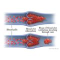
A Doppler ultrasound, Opens dialog test uses reflected sound waves to see how blood flows through a blood vessel. It helps doctors assess the blood flow through major arteries and veins, such as those of the arms, legs, and neck. It can show blocked or reduced flow of blood through narrow areas in the major arteries of the neck. This problem could cause a stroke, Opens dialog. The test also can find blood clots in leg veins (deep vein thrombosis, or DVT, Opens dialog) that could break loose and block blood flow, Opens dialog to the lungs. This problem is called a pulmonary embolism. During pregnancy, Doppler ultrasound may be used to look at blood flow in an unborn baby to check the baby's health.
During Doppler ultrasound, Opens dialog, a handheld device is passed lightly over the skin above a blood vessel. The device is called a transducer. It sends and receives sound waves that are amplified through a microphone. The sound waves bounce off solid objects, including blood cells. The movement of blood cells causes a change in the pitch of the reflected sound waves. This is called the Doppler effect. If there is no blood flow, the pitch does not change.
Information from the reflected sound waves can be used to make graphs or pictures that show the flow of blood through the blood vessels. These graphs or pictures can be saved and reviewed later.
The three basic types of Doppler ultrasound are:
- "Bedside" or continuous wave Doppler.
- This type uses the change in pitch of the sound waves to provide information about blood flow through a blood vessel. The doctor listens to the sounds made by the transducer to assess the blood flow through an area that may be blocked or narrowed. This type of ultrasound can be done at the bedside in the hospital. It uses a portable machine that can quickly check the extent of blood vessel damage or disease.
- Duplex Doppler.
- This test uses standard ultrasound methods to make a picture of a blood vessel and the organs around it. A computer turns the Doppler sounds into a graph. This graph helps to show the speed and direction of blood flow through the blood vessel.
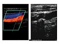
- Color Doppler.
- Color Doppler uses standard ultrasound methods to make a picture of a blood vessel. A computer changes the Doppler sounds into colors that are overlaid on the image of the blood vessel. These colors show the speed and direction of blood flow through the vessel. Power Doppler is a special type of color Doppler. Power Doppler can get some images that are hard or impossible to get using standard color Doppler. Power Doppler is most often used to look at blood flow through vessels within solid organs.
This test is done in an ultrasound room in a hospital or doctor's office.
You will need to remove any jewelry that might affect the Doppler ultrasound scan. You may be asked to drink water right before the test so that your bladder is full. You may need to take off all or most of your clothes, depending on which part of the body is being examined. You may be allowed to keep on your underwear if it does not affect the test. You will be given a cloth or paper covering to use during the test.
- For abdominal scans, you will lie on your back.
- For chest scans, you will lie on your back with your neck slightly extended.
- For head and neck scans, your head may be turned to one side.
- For an arm or leg scan, your head will be slightly raised. The exposed arm or leg will be turned slightly outward. Sometimes for a leg scan, you may be asked to lie on your stomach.
- During pregnancy, you will lie on your back or on your left side with your belly exposed.
Gel is applied to the skin to help the sound waves pass through. The transducer is placed in the gel and moved along the skin. You need to lie very still during the test. You may hear sounds from the flow of blood through the blood vessels.
Arteries in the arms and legs
This test is often done on both arms or both legs. Even if the suspected blood flow problem is in only one limb, both may be tested to compare them. If your arms are being tested, they will be tested first while you lie down. Then they'll be tested again while you sit.
Depending on which blood vessels are being tested, a blood pressure cuff may be wrapped around one or both limbs. It allows your blood pressure to be taken at several different places. When the legs are tested, a blood pressure cuff may be wrapped first around the calf and then around the thigh. The test may be done at several places on your leg. When the arms are tested, the pressure cuff may be wrapped first around the forearm and then around the upper arm.
Testing may be done before and after exercise, if you are healthy enough.
Veins in the arms and legs
For this test, you will be asked to lie down and breathe normally. You must lie very still. Any changes in blood flow that are affected by how you breathe are noted.
The test may be repeated while the examiner presses on the veins close to the surface of your skin. This helps to find a clot in the vein. It's called a compression maneuver. The examiner may do this maneuver with your legs or arms in different positions. This is to make sure that the blood supply is not blocked in these positions. He or she may also squeeze your calf or forearm to help blood move more quickly through the veins. This is called an augmentation maneuver. It is done to check blood flow toward your heart.
While your legs are being tested, you may also be asked to try to breathe out strongly with your nose pinched and your mouth closed. This is called a Valsalva maneuver, Opens dialog. It usually causes a sudden change in blood flow through the veins.
Arteries in the neck
You will be asked to lie down with a pillow under your head for support. The test is done on both sides of your neck. Then the results are compared to standard values to find out how much the arteries are blocked or narrowed.
Transcranial ultrasound
For this type of ultrasound, the transducer is passed lightly over the skin at the base or side of your skull.
During pregnancy
The transducer is moved back and forth on your belly until the doctor finds the blood vessel that needs to be studied. After the doctor has found the blood vessel, it may take some time to assess the blood flow.
How long the test takes
The test will take 30 to 60 minutes.
This test usually doesn't cause any pain or discomfort.
There are no known risks from having this test.
The scans from the test will be read within a short time.
Doppler ultrasound Normal: | The test does not show significant narrowing or other problems in any of the arteries. |
|---|
| There is no sign of a clot in any of the veins examined. The size and position of veins are normal. |
| Normal blood flow is found in the blood vessels that supply oxygen and nutrients to a fetus, Opens dialog. |
Abnormal: | For continuous wave Doppler or duplex Doppler, differences in blood flow between the right and left sides of the body may be heard. At the exact spot where an artery is blocked or narrowed, the sound may be high-pitched or turbulent. Blockage (such as from a blood clot), an aneurysm, Opens dialog, or narrowing of a blood vessel may be found. The speed of blood flow may be compared to standard values to find out how blocked or narrow the blood vessel is. |
|---|
| A duplex Doppler ultrasound graph may show blood flow that isn't normal. This is a sign of a blocked or narrowed blood vessel. |
| A color Doppler image may show a blocked or narrowed blood vessel or an aneurysm. |
| In the veins, a blood clot may be present if blood flow does not change in response to breathing or does not increase after either a compression maneuver or Valsalva maneuver, Opens dialog. Incomplete blockage of a vein by a blood clot may be seen on color Doppler or during a compression maneuver. |
| Abnormal veins, such as varicose veins, Opens dialog, are seen. |
| There is an abnormal increase or decrease in blood flow through the vessels that supply oxygen and nutrients to a fetus. |