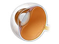
The slit lamp exam uses a tool that provides a magnified, three-dimensional (3-D) view of the parts of the eye, Opens dialog. During the exam, your doctor can look at the front parts of the eye. These parts include the clear, outer covering (cornea), the lens, and the colored part (iris). The doctor can also see the front part of the thick fluid (vitreous gel) that fills the large space in the middle of the eye.
Special lenses can be placed between the slit lamp and the cornea (or on the cornea) to help the doctor see the deeper structures of the eye. These structures include the optic nerve, the retina, and the area where fluid drains out of the eye (drainage angle, Opens dialog).
A camera may be attached to the slit lamp to take pictures of different parts of the eye.
Fluorescein dye may be used during a slit lamp exam. The dye makes it easier to see a foreign object, such as a metal fragment, or an infected or injured area on the cornea.
Routine slit lamp exams are done to find eye problems at an early stage and to guide treatment if eye problems develop.
A slit lamp exam may be done:
As part of a routine eye exam. It may be used along with other procedures that evaluate the eye, such as ophthalmoscopy, vision testing, and tonometry (to measure pressure in the eye).
To look at structures in the back of the eye, such as the optic nerve or retina.
To keep checking on problems such as bleeding after an eye injury.
To keep checking on problems such as cataracts that form because of chemotherapy or radiation treatment or after a bone marrow transplant.
If you wear glasses or contact lenses, you will need to remove them before the slit lamp exam.
Eyedrops may be used to widen (dilate) your pupils, Opens dialog and to numb the surface of your eyes. Before the test, tell your doctor if you have glaucoma or are allergic to eyedrops that dilate or numb your eyes.
If dilating drops are used, your eyes may be sensitive to light. You will have trouble focusing your eyes for several hours. If you know that your eyes will be dilated, you may wish to find someone to drive you home after the test. You also will need to wear sunglasses when you go outside or into a brightly lit room.
The doctor may put one or more types of drops in your eye. Dilating drops may be used to make the opening (pupil) in the center of the eye bigger. This makes it easier for the doctor to see the structures of your eye. Numbing drops may be used if a foreign object is to be removed or if eye pressure is being checked (tonometry). In some cases, fluorescein dye is used.
You will sit in a chair and rest your chin and forehead against bars on the slit lamp. The lights in the room will be dimmed.
The slit lamp will be placed in front of your eyes, in line with the doctor's eyes. Focus your eyes in the direction the doctor tells you to. Try to hold your eyes steady without blinking.
A narrow beam of bright light from the slit lamp is directed into your eye while the doctor looks through the microscope. In some cases, a camera may be attached to the slit lamp to take pictures of different parts of the eye.
A test called fluorescein staining may be done along with a slit lamp exam.
During this test, your doctor applies a dye called fluorescein. The dye comes in an eyedrop or as a paper strip that is gently touched to the inside of your lower eyelid. The dye dissolves in your tears, coats your cornea, and collects for a short time in any scratches or other abnormal areas. The rest of the dye is washed away by your tears.
Your doctor shines a light onto your eye. The fluorescein dye shows up under the light. It helps the doctor to see scratches, ulcers, burns, or areas of irritation from an infection or dryness.
A slit lamp exam takes about 5 to 10 minutes.
It is usually not painful to have a slit lamp test.
The dilating drops may make your eyes sting and cause a medicine taste in your mouth. You will have trouble focusing your eyes for up to 12 hours. Your distance vision usually is not affected as much as your near vision. But your eyes may be very sensitive to light. Do not drive for several hours after your eyes have been dilated, unless your doctor says it's okay. Wearing sunglasses may make you more comfortable until the effect of the drops wears off.
Numbing drops usually wear off in about 30 minutes.
In some people, the dilating or numbing drops can cause:
Some nausea, vomiting, dryness of the mouth, flushing, and dizziness for a short time.
Contact your doctor right away if you have severe and sudden eye pain, vision problems such as halos that appear around lights, or loss of vision after the exam.
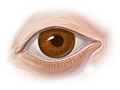
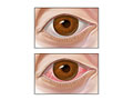
Slit lamp exam
Normal:
The eyelashes, eyelids, and lining of the eyelids (conjunctiva) look normal.
Author: Ignite Healthwise, LLC Staff Clinical Review Board All Ignite Healthwise, LLC education is reviewed by a team that includes physicians, nurses, advanced practitioners, registered dieticians, and other healthcare professionals.
Clinical Review Board All Ignite Healthwise, LLC education is reviewed by a team that includes physicians, nurses, advanced practitioners, registered dieticians, and other healthcare professionals.
This information does not replace the advice of a doctor. Ignite Healthwise, LLC, disclaims any warranty or liability for your use of this information. Your use of this information means that you agree to the Terms of Use. Learn how we develop our content.
This information does not replace the advice of a doctor. Ignite Healthwise, LLC, disclaims any warranty or liability for your use of this information. Your use of this information means that you agree to the Terms of Use. Learn how we develop our content.
To learn more about Ignite Healthwise, LLC, visit webmdignite.com.