Spinal X-rays are pictures of the spine. They may be taken to find injuries or diseases that affect the discs, Opens dialog or joints in your spine. These problems may include spinal fractures, infections, dislocations, tumors, bone spurs, or disc disease.
Spinal X-rays are also done to check the curve of your spine (scoliosis, Opens dialog) or for spinal defects.
The spine is divided into four parts. So there are four common types of spinal X-rays:
Cervical spine X-ray.
It takes pictures of the 7 neck (cervical) bones.
Thoracic spine X-ray.
It takes pictures of the 12 chest (thoracic) bones.
Lumbosacral spine X-ray.
It takes pictures of the 5 bones of the lower back (lumbar vertebrae) and a view of the 5 fused bones at the bottom of the spine (sacrum).
Sacrum/coccyx X-ray.
It takes a detailed view of the 5 fused bones at the bottom of the spine (sacrum) and the 4 small bones of the tailbone (coccyx).
The most common spinal X-rays are of the cervical vertebrae (C-spine films) and lumbosacral vertebrae (LS-spine films).
Check the spine for effects from other problems, such as infections, tumors, or bone spurs.
Check for abnormal curves of the spine, such as scoliosis, in children or young adults.
Check the spine for problems present at birth (congenital conditions), such as spina bifida, Opens dialog, in infants, children, or young adults.
Check changes in the spine after spinal surgery.
In general, there's nothing you have to do before this test, unless your doctor tells you to.
Information about Spinal X-Ray
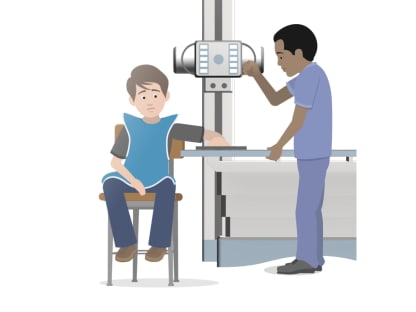
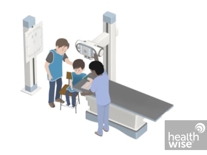
You will need to remove any jewelry that may be in the way of the X-ray picture. You may need to take off some of your clothes, depending on which area is examined. You will be given a cloth or paper gown to use during the test. You may be allowed to keep on your underwear if it does not get in the way of the test.
During the X-ray test, you will lie on an X-ray table. If the X-ray is being taken because of a possibly serious injury to your neck or back, a radiologist will look at the first X-ray pictures before taking others. This is done to prevent causing more injury. If you have a neck brace (cervical collar) in place, X-ray pictures may be taken and a physical exam done to see whether the brace can be taken off without hurting the spine.
Usually 3 to 5 X-ray pictures are taken. You need to lie very still to avoid blurring the pictures.
How long the test takes
A spinal X-ray usually takes about 15 minutes. You will wait about 5 minutes until the X-rays are processed in case more pictures need to be taken. In some clinics and hospitals, X-ray pictures can be shown right away on a computer screen.
You won't feel any pain from the X-ray itself. You may find that the positions you need to hold are uncomfortable or painful. This is more likely if you have an injury.
There is always a slight chance of damage to cells or tissue from radiation, including the low levels of radiation used for this test. But the chance of damage from the X-rays is extremely low. It is not a reason to avoid the test.
If you need an X-ray during pregnancy, a lead apron will be put over your belly to protect the baby from exposure to radiation from the X-rays. The chance of harm is usually very low compared with the benefits of the test.
In an emergency, a doctor can see the results of a spinal X-ray in a few minutes. Otherwise, a radiologist, Opens dialog usually has the official X-ray report ready the next day.
Spinal X-ray
Normal:
The bones of the spine (vertebrae) are normal in number, size, shape, appearance, and how they are lined up.
No broken bones, dislocations, or foreign objects are present. The soft tissues around the vertebrae look normal.
The spine is not abnormally curved.
Abnormal:
Broken bones, dislocations, or foreign objects are present.
Diseases that affect the spine, such as thin bones (osteoporosis, Opens dialog) or arthritis, Opens dialog, are present. One or more bones in the spine may be abnormal because of a condition you were born with or because of cancer, infection, or trauma.
Disc disease, which is fairly common, can sometimes be seen on a spinal X-ray as a narrowed space between the bones of the spine. Bone spurs can also be seen.
Author: Ignite Healthwise, LLC Staff Clinical Review Board All Ignite Healthwise, LLC education is reviewed by a team that includes physicians, nurses, advanced practitioners, registered dieticians, and other healthcare professionals.
Clinical Review Board All Ignite Healthwise, LLC education is reviewed by a team that includes physicians, nurses, advanced practitioners, registered dieticians, and other healthcare professionals.
This information does not replace the advice of a doctor. Ignite Healthwise, LLC, disclaims any warranty or liability for your use of this information. Your use of this information means that you agree to the Terms of Use. Learn how we develop our content.
This information does not replace the advice of a doctor. Ignite Healthwise, LLC, disclaims any warranty or liability for your use of this information. Your use of this information means that you agree to the Terms of Use. Learn how we develop our content.
To learn more about Ignite Healthwise, LLC, visit webmdignite.com.